Testicular Ultrasound

Ultrasound imaging of the scrotum is the primary imaging method used to evaluate disorders of the testicles.

This study is typically used to help diagnose symptoms such as:

  • a mass in the scrotum felt by the patient or doctor
  • trauma to the scrotal area
  • testicular pain or swelling

Ultrasound is also a valuable tool for evaluating the epididymis (a tube that collects sperm made by the testicles) and the prostate.


Some of the problems ultrasound imaging can identify include:

  • inflammation of the scrotum
  • Collection of Fluid i.e hydrocele
  • Varicose veins
  • an absent or undescended testicle
  • testicular torsion
  • abnormal blood vessels
  • a lump or tumor

Thyroid Ultrasound

An ultrasound of the thyroid produces a picture of the thyroid gland. The thyroid gland is located in front of the neck just below the Adam’s apple and is shaped like a butterfly, with two lobes on either side of the neck connected by a narrow band of tissue. It is one of nine endocrine glands located throughout the body that make and send hormones into the bloodstream.
 
An ultrasound of the thyroid is typically used to help diagnose:

  • A lump in the thyroid
  • A thyroid that is not functioning properly.
  • Because ultrasound provides real-time images, it also can be used to guide procedures such as needle biopsies, in which needles are used to extract sample cells from an abnormal area for laboratory testing.

Renal Ultrasound

The kidneys are a pair of bean-shaped organs located toward the back of the abdominal cavity, just above the waist. They remove waste products from the blood and produce urine. The ureters are thin tubes that carry the urine from the kidneys to the bladder.


A renal ultrasound is a safe and painless test that uses sound waves to make images of the kidneys, ureters, and bladder.


Renal ultrasound tests can show:

  • the size of the kidneys
  • signs of injury to the kidneys
  • abnormalities present since birth
  • the presence of blockages or kidney stones
  • complications of a Urinary tract infection.
  • cysts or tumors

Pelvic Ultrasound

A Pelvic Ultrasound looks at:

  • The bladder, ovaries, uterus, cervix, and fallopian tubes of a woman.
  • The bladder, prostate gland, and seminal vesicles of a man.

Organs and structures that are solid and uniform, like the uterus, ovaries, or prostate gland, or are fluid-filled, like the bladder, show up clearly on a pelvic ultrasound.


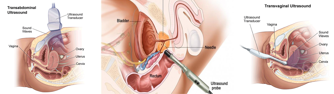
Pelvic ultrasound can be done three ways: transabdominal, transrectal, and transvaginal.

Transabdominal ultrasound.

A small handheld device called a transducer is passed back and forth over the lower belly. A transabdominal ultrasound is commonly done in women to look for large uterine fibroids or other problems.

Transrectal ultrasound.

The transducer is shaped to fit into the rectum. A transrectal ultrasound is the most common test to look at the male pelvic organs, such as the prostate and seminal vesicles. Sometimes, a small sample of tissue (biopsy) may be taken with small tools inserted through the rectum during a transrectal ultrasound.

Transvaginal ultrasound.

The transducer is shaped to fit into a woman’s vagina. A woman may have both transabdominal and transvaginal ultrasounds to look at the whole pelvic area. A transvaginal ultrasound is done to look for problems with fertility. In rare cases, a hysterosonogram is done to look at the inside of the uterus by filling the uterus with fluid during a transvaginal ultrasound. Sometimes, a small sample of tissue (biopsy) may be taken with small tools inserted through the vagina during a transvaginal ultrasound.

Abdominal Ultrasound

Indications for an ultrasound examination of the abdomen and/or retroperitoneum includes as follows:

  • Abdominal, flank, and/or back pain.
  • Signs or symptoms that may be referred from the abdominal and/or retroperitoneal regions, such as jaundice or hematuria.
  • Palpable abnormalities such as an abdominal mass or organomegaly.
  • Abnormal laboratory values or abnormal findings on other imaging examinations suggestive of abdominal and/or retroperitoneal pathology.
  • Follow-up of known or suspected abnormalities in the abdomen and/or retroperitoneum.
  • Search for metastatic disease or an occult primary neoplasm.
  • Evaluation of cirrhosis, portal hypertension, and transjugular intrahepatic portosystemic shunt (TIPS) stents; screening for hepatoma; and evaluation of the liver in conjunction with liver elastography.
  • Abdominal trauma.
  • Evaluation of urinary tract infection and hydronephrosis.
  • Evaluation of uncontrolled hypertension and suspected renal artery stenosis.
  • Search for the presence of free or loculated peritoneal and/or retroperitoneal fluid.
  • Evaluation of suspected congenital abnormalities.
  • Evaluation of suspected hypertrophic pyloric stenosis, intussusception, necrotizing enterocolitis, or any other bowel abnormalities.
  • Pre-transplantation and post-transplantation evaluation.
  • Planning for and guiding an invasive procedure.

Aorta Duplex Ultrasound

Indications for ultrasound of the abdominal aorta include:

A. Diagnostic Evaluation for an Abdominal Aortic Aneurysm

  • Palpable or pulsatile abdominal mass or abdominal bruit
  • Unexplained lower back pain, flank pain, or abdominal pain.
  • Follow-up of a previously demonstrated abdominal aortic aneurysm.
  • Follow-up of patients with an abdominal aortic and/or iliac endoluminal stent graft.

B. Screening Evaluation for an Abdominal Aortic Aneurysm

  • Men 65 years or older.
  • Women 65 years or older with cardiovascular risk factors.
  • Patients 50 years or older with a family history of aortic and/or peripheral vascular aneurysmal disease.
  • Patients with a personal history of peripheral vascular aneurysmal disease

Renal Artery Ultrasound

Renal Arteries provide blood flow to the kidneys.


Indications for renal artery duplex sonography include to:

  • Evaluation of patients with hypertension, particularly when there is a moderate to high suspicion of renovascular hypertension.
  • Follow-up of patients with known renovascular disease who have undergone renal artery stent placement or other renal artery interventions or have known unilateral stenosis with concern for stenosis in the contralateral kidney;
  • Evaluation of an abdominal or flank bruit;
  • Evaluation of a suspected vascular abnormality such as an aneurysm, pseudoaneurysm, arteriovenous malformation, or arteriovenous fistula;
  • Evaluation of renal insufficiency in a patient at risk for renovascular disease.
  • Evaluation of renal artery blood flow in patients with known aortic dissection, trauma, or other abnormalities that may compromise blood flow to the kidneys.
  • Evaluation of discrepant renal size.

Venous Duplex Ultrasound

The indications for peripheral venous ultrasound examinations include :

The indications for peripheral venous ultrasound examinations include :

  • Evaluation of possible venous thromboembolic disease or venous obstruction in symptomatic or high-risk asymptomatic individuals.
  • Serial evaluation may be necessary in some high-risk individuals (eg, based on history, pretest probability, and/or D-dimer test) whose initial examination is negative for deep venous thrombosis.
  • Assessment of venous insufficiency, reflux, and varicosities.
  • Postprocedural assessment of venous ablation or other interventions.
  • Assessment of dialysis access.
  • Venous mapping before surgical procedure.
  • Evaluation of veins before venous access.
  • Follow-up for patients with known venous thrombosis at or near the anticipated end of anticoagulation to determine if residual venous thrombosis is present.

Arterial Duplex Ultrasound

Duplex ultrasonography of the Peripheral arteries (upper and lower ) is performed to provide an overview of the location, extent and severity of vascular disease which includes :

  • Detection of stenosis or occlusions in segments of the peripheral arteries in symptomatic patients with suspected arterial occlusive disease. These patients could present with claudication, rest pain, ischemic tissue loss, an aneurysm, and arterial embolization.
  • Monitoring of sites of previous surgical interventions, including sites of previous bypass surgery with either synthetic or autologous vein grafts.
  • Monitoring of sites of various percutaneous interventions, including angioplasty, thrombolysis/ thrombectomy, atherectomy, and stent placements.
  • Follow-up for progression of previously identified disease, such as documented stenosis in an artery that has not undergone intervention, aneurysms, atherosclerosis, or other occlusive diseases.
  • Evaluation of suspected vascular and perivascular abnormalities, including such entities as masses, aneurysms, pseudoaneurysms, arterial dissections, vascular injuries, arteriovenous fistulas, thromboses, emboli, and vascular malformations.
  • Mapping of arteries before surgical interventions.
  • Clarifying or confirming the presence of significant arterial abnormalities identified by other imaging modalities.
  • Evaluation of arterial integrity in the setting of trauma.
  • Evaluation of patients suspected of thoracic outlet syndrome, such as those with positional numbness, pain tingling, or a cold hand.
  • The Allen test to establish patency of the palmar arch.

Transcranial Doppler

Transcranial Doppler (TCD) is a test that measures the velocity of blood flow through the brain’s blood vessels.
 
We perform a comprehensive and detailed protocol for understanding and interrogating a wide variety of neurovascular pathology such as:

  • Detecting Intracranial Stenosis and occlusions.
  • Assess collateral circulation.
  • Cerebral Emboli Detection.
  • Evaluate onset and severity of Cerebral Vasospasm.
  • Evaluate intracranial Arteriovenous Malformations
  • Variations in Circle of Willis.